Chorionic Bump in Early Pregnancy
Department of Radiodiagnosis
Aadhar Health Institute, Hisar
Consultants
Dr. Priyanka Goyal, Dr. Pooja Batra, Dr. Lovekesh Bhatia, Dr. Shweta,
Residents
Dr. Raveesh, Dr. Kshitijaa, Dr. Neetika, Dr. Priya
Case Introduction
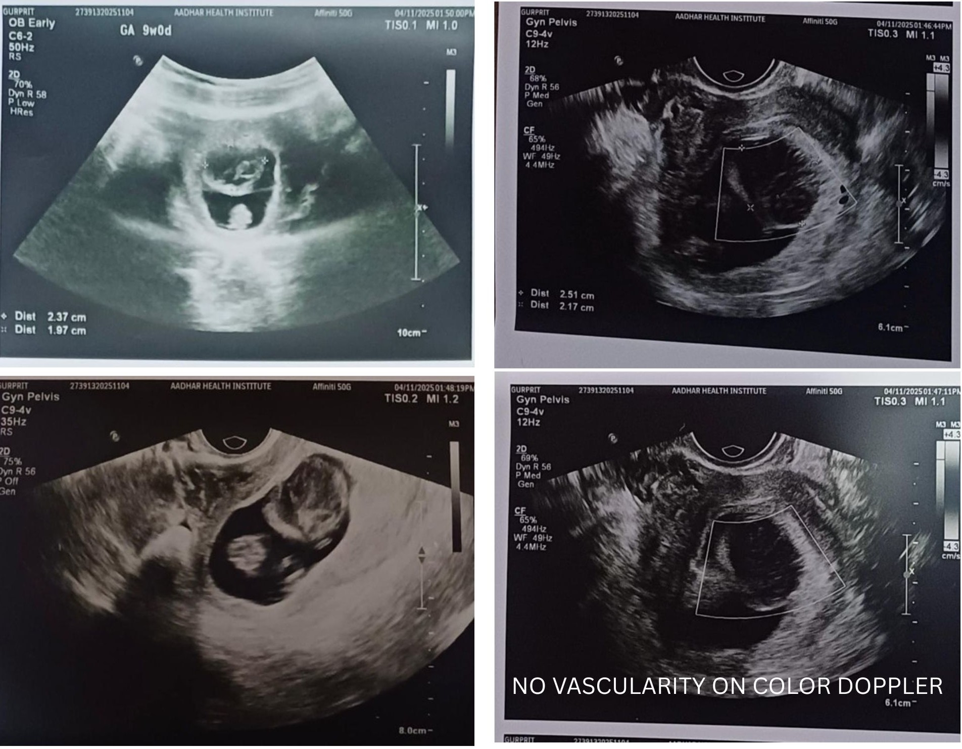
A 28-year-old female presented for a routine early pregnancy ultrasound at 9 weeks of gestation.
USG Images
.jpg)
.jpg)
Ultrasound Findings
-
Single live intrauterine pregnancy
-
Cardiac activity present
-
A well-marginated heteroechoic lesion projecting into the gestational sac from the chorionic wall
-
No internal vascularity on Color Doppler
-
No subchorionic hematoma or free fluid
.jpg)
Introduction
A chorionic bump is a focal, convex, nodular bulge arising from the chorionic surface and projecting into the gestational sac. It usually shows mixed or hypoechoic internal echoes.
It may represent a small hematoma bulging into the gestational sac or a resorbing anembryonic (failed) second gestation. It is an uncommon ultrasound finding, seen in approximately 0.4–0.7% of pregnancies before 10 weeks.
Pathology
Primary hypothesis:
-
Focal hematoma within the chorionic plate causing outward bulging into the gestational sac
Alternative theories:
-
Abnormal placentation or implantation site reaction
-
Local vascular malformation or thrombosis within chorionic villi
-
Focal decidual necrosis due to poor perfusion
-
Area of early chorionic injury compromising trophoblastic function if extensive
Radiological Features
Ultrasound:
-
Irregular, convex soft-tissue bulge projecting from the choriodecidual surface
-
Isoechoic or heteroechoic compared to chorion
-
Avascular on Color Doppler
-
No associated subchorionic hematoma
MRI:
-
May show T1 hyperintensity suggesting hemorrhagic content
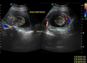
Differential Diagnosis
| Entity | Key Imaging Features |
|---|---|
| Chorionic bump | Localized convex echogenic/heteroechoic bulge projecting into gestational sac; avascular |
| Subchorionic hematoma | Crescent-shaped hypoechoic collection peripheral to gestational sac |
| Decidual reaction | Uniform, symmetrical echogenic thickening around gestational sac |
| Embryonic/Fetal pole | Definite structure with cardiac activity |
| Decidual cyst | Small anechoic cystic lesion within decidua |
Treatment & Prognosis
No specific treatment is required; close sonographic follow-up is recommended.
Chorionic bump is associated with a guarded prognosis in early pregnancy. The risk of miscarriage is approximately twice that of normal pregnancies.
Studies report a 62–65% overall live birth rate, improving to approximately 83% in the absence of other abnormalities. Multiple chorionic bumps are associated with poor outcomes, with reported fetal loss approaching 100%.