Multidisciplinary Management of Multi-Focal Hepatic Hydatid Cysts with Post-Operative ERCP Evaluation
Executive Summary: Hydatid disease (Echinococcosis) involving central liver segments presents significant clinical challenges due to the risk of cysto-biliary communications. This report details the successful surgical management and subsequent post-operative endoscopic evaluation (ERCP with CBD stenting) in a patient with multiple hepatic hydatid cysts, confirming biliary tree integrity and facilitating uneventful recovery.
1. Patient Presentation & Initial Workup
A patient presented for surgical evaluation with findings indicative of hepatic space-occupying lesions. Comprehensive baseline laboratory evaluation and radiological imaging were performed to establish the diagnosis and assess biliary anatomy.
Baseline Laboratory Profile (06/08/2026)
| Parameter | Observed Value | Reference / Units |
|---|---|---|
| Hemoglobin (Hb) | 13.10 g/dL | 13.0 – 17.0 g/dL |
| Total Leukocyte Count (TLC) | 9.7 × 10³/µL | 4.0 – 11.0 × 10³/µL |
| Platelet Count | 235 × 103/µL | 150 – 450 × 103/µL |
| Serum Bilirubin (Total) | 1.01 mg/dL | 0.2 – 1.2 mg/dL |
| SGPT (ALT) / SGOT (AST) | 985 / 1089 U/L | Elevated (Transaminitis) |
| Alkaline Phosphatase (ALP) | 181 U/L | 40 – 129 U/L |
| Serum Creatinine / Albumin | 0.64 mg/dL / 4.53 g/dL | Normal limits |
| Viral Markers (HIV, HCV, HBsAg) | Non-Reactive | Negative |
2. Radiological Evaluation
Contrast-Enhanced CT (CECT) Whole Abdomen (06/08/2026)
The CECT scan revealed a normal-sized liver with non-dilated intrahepatic biliary radicles (IHBRs). Three distinct, well-defined lesions were identified within the hepatic parenchyma:
- Lesion 1 (Segment IVB): Measures 65 × 61 × 52 mm.
- Lesion 2 (Segment VII/VIII): Measures 48 × 39 × 42 mm.
- Lesion 3 (Segment II): Measures 18 × 19 mm.
Ultrasonography (USG) Upper Abdomen (07/08/2026)
Ultrasound confirmed a liver span of 15.6 cm and further characterized the structural composition of the primary lesions:
- Segment IV Complex Cyst: Cystic lesion containing daughter cysts measuring 4.8 × 5.7 × 7.7 cm, bulging prominently into the portal region and indenting the left hepatic duct including the primary biliary confluence.
- Segment VII Complex Cyst: Complex cyst with internal septations and peripheral daughter cysts measuring 4.5 × 4.9 × 4.5 cm.
3. Operative & Post-Operative Course
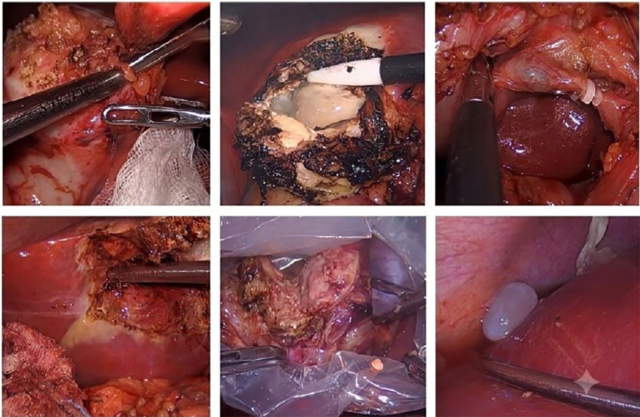
The patient underwent definitive surgical management consisting of cyst deroofing, scolicidal agent instillation, evacuation of daughter cysts, and placement of intra-abdominal drains. Post-operatively, serial laboratory monitoring on 09/08/2026 revealed transient hyperbilirubinemia (Total Bilirubin: 3.09 mg/dL) and elevated inflammatory markers (TLC: 16.0 × 10³/μL, SGPT/SGOT: 761 / 337 U/L).
4. Endoscopic Retrograde Cholangiopancreatography (ERCP)
In view of the anatomic compression on the left hepatic duct and primary confluence demonstrated preoperatively, a diagnostic and therapeutic ERCP was undertaken to rule out cysto-biliary fistula or active leak.
- Cholangiography Findings: Selective cannulation confirmed normal caliber major bile ducts with no evidence of contrast extravasation or active bile leak.
- Intervention: A therapeutic Common Bile Duct (CBD) stent was successfully deployed to maintain biliary decompression, minimize intraductal pressure, and ensure safe postoperative healing.
5. Patient Outcome & Clinical Takeaway
Following CBD stenting, the patient made a smooth recovery. Abdominal drain output remained minimal and nonbilious. On 17/08/2026, the patient was discharged in a Medically Fit, stable condition with the abdominal drain remaining in situ for outpatient follow-up. Conclusion: In centrally located hepatic hydatid cysts close to the primary biliary confluence, combining meticulous surgical evacuation with proactive post-operative ERCP stenting safely rules out occult biliary leaks and prevents high-pressure biliary complications.