
Sinonasal Adenocarcinoma – Extensive Right Nasal Cavity Tumour
CLINICAL PRESENTATION
Presented with a right nasal cavity mass with bilateral nasal obstruction and headache. Clinical assessment documented a right-sided nasal cavity mass; endoscopic excision was planned.
PRE-OPERATIVE IMAGING
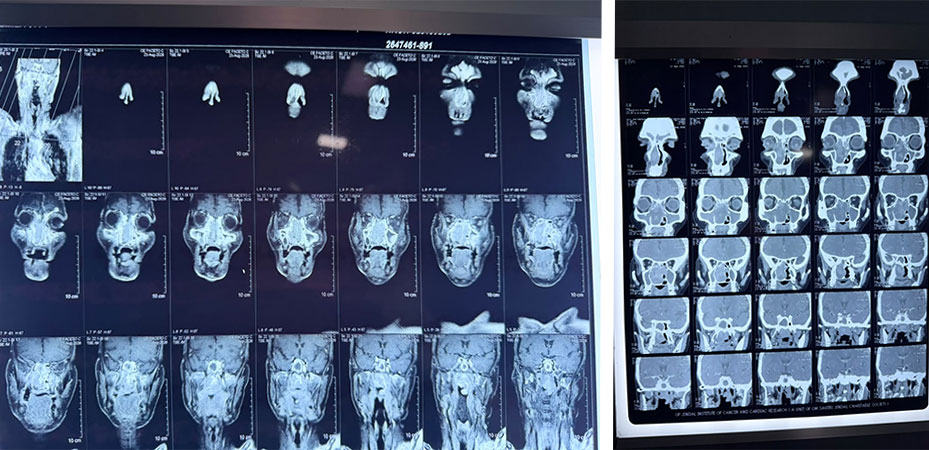
CECT PNS showed a large enhancing soft-tissue lesion occupying and expanding the right nasal cavity and ethmoid air cells, with leftward septal displacement, right OMC obstruction, thinning of the right lamina papyracea with orbital bulging, superior ethmoid extension and posterior extension toward the nasopharynx.
MRI demonstrated a polypoidal right nasal cavity mass measuring approximately 8.6 × 3.8 cm, extending to the right OMC/posterior choana, with septal displacement and erosion of the right lamina papyracea and ethmoid air cells. No intracranial or intraoral extension was reported.
HISTOPATHOLOGY
Biopsy from the right nasal cavity showed glandular and papillary structures with mild-to-moderate nuclear pleomorphism/hyperchromasia and focal necrosis. Diagnosis: SINONASAL ADENOCARCINOMA.
SURGICAL MANAGEMENT
Endoscopic En-bloc Excision of Extensive Sinonasal Tumour
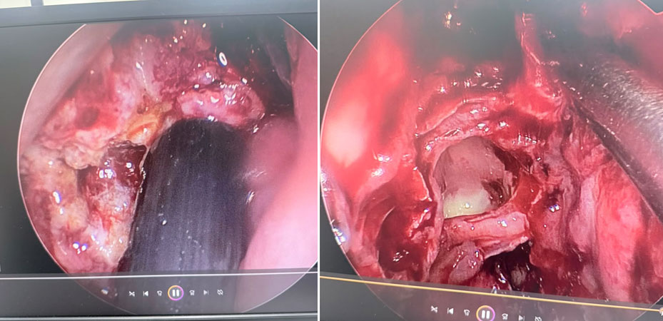
A completely endoscopic approach was used for management of the extensive right nasal cavity tumour, allowing wide surgical access without any external incision.
- Medially, the tumour was detached en bloc along with the right nasal septal mucosa and underlying septal cartilage, which was taken as a margin.
- Laterally, the tumour was separated from the maxillary sinus using a modified Denker’s approach, providing adequate access to the lateral extent of the lesion.
- Superiorly, the tumour was meticulously detached from the anterior and posterior ethmoid regions. The lesion had superior extension up to the skull base, requiring careful endoscopic dissection with preservation of the relevant skull-base landmarks.
- After complete circumferential mobilization, the tumour was delivered posteriorly into the nasopharynx.
- The entire tumour was subsequently removed in toto through the oral cavity, avoiding fragmentation of the specimen.
- Throughout the procedure, all major anatomical landmarks and critical structures were identified and preserved.
- The procedure was completed entirely endoscopically with no external incision or visible facial scar, resulting in a scarless surgical approach.
KEY LEARNING POINT
A unilateral nasal mass with heterogeneous enhancement and adjacent bone erosion/remodelling should prompt early imaging and tissue diagnosis, with careful assessment of orbital, skull-base and nasopharyngeal relationships before definitive surgery.
Dr Dishant Chhabra, MS ENT, FHNS
Consultant ENT & Head & Neck Oncosurgeon
Aadhar Health Institute, Hisar