When MRI Doesn’t Tell the Whole Story: A Rare Presentation of AQP4-IgG Positive NMOSD
When MRI Doesn’t Tell the Whole Story: A Rare Presentation of AQP4-IgG Positive NMOSD
Dr. Ramnik Garg
Consultant Neurologist, Aadhar Health Institute, Hisar
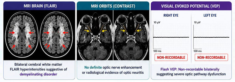
A 54-year-old woman presented with rapidly progressive, painless bilateral vision loss over 10 days. Ophthalmological examination was unremarkable. MRI brain showed bilateral white matter demyelinating lesions, while contrast MRI of the orbits revealed no evidence of optic neuritis.

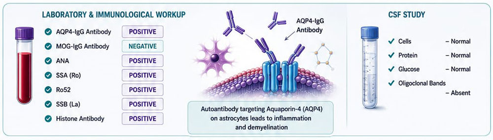
Further evaluation demonstrated positive AQP4-IgG antibodies, negative MOG-IgG, normal CSF with absent oligoclonal bands, and positive ANA, SSA (Ro), Ro52, SSB (La), and histone antibodies. Flash visual evoked potentials were non-recordable bilaterally, confirming severe optic pathway involvement despite a normal orbital MRI.
She was treated with intravenous methylprednisolone, followed by five sessions of plasma exchange and Rituximab, resulting in significant visual improvement.
Key Message:
A normal orbital MRI does not exclude NMOSD-related optic neuritis. In patients with acute bilateral vision loss, AQP4-IgG testing and visual evoked potentials are crucial for diagnosis. Early immunotherapy can be vision-saving.